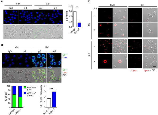
Figure 5.
α-TREM-1 promotes bacterial clearance through increased macrophage autophagy. (A,B) RAW264.7 cells were pre-treated with IgG or α-TREM-1 and infected with S. typhimurium expressing green-fluorescent protein (A, multiplicity of infection MOI = 10; B, MOI = 100) for 1 h. (A) Representative images of S. typhimurium-GFP-infected macrophages and quantification of the total number of bacteria per macrophage (right). Scale bar, 20 μm. (B) Representative images of lysotracker (Lyso)-stained macrophages and the percentage of S. typhimurium-GFP in autophagic degradation (GFP co-localization with lysosomes). GFP+Lyso+ area was calculated by subtracting pure green GFP signal from total GFP signal. Scale bar, 20 μm. Hoechst (Hoec) staining of nuclei. (C) Representative images of lysotracker-stained macrophages. Scale bar, 40 μm. RAW264.7 cells transfected with scrambled (SCR) or TREM1-specific (siT) siRNA were stimulated with LPS for 4 h after pre-treatment with α-TREM-1. Data are expressed as means ± S.E.M. of at least three independent experiments. Statistical significance was assessed using Student t-test. *P < 0.05, ***P < 0.005. α-T, treated with α-TREM-1; DIC, differential interference contrast; IgG, treated with control antibody; Sal, infected with S. typhimurium-GFP; Veh, treated with vehicle.

