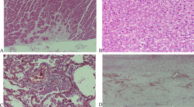
Fig. 6.

Case #3. A Focal area of myocardial scar (H&E, × 200). B Steatosis of Hepatocytes (H&E, × 200). C Patchy lymphocytic interstitial infiltration and fibrinous exudate in the alveoli. This image was taken from one of the few areas where inflammation was obvious. In other areas, infiltration was sparse or absent (H&E, × 400). D Organizing subdural hemorrhage (H&E, × 200)
