FIGURE 3.
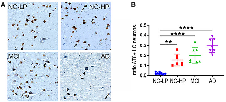
Locus coeruleus (LC) tau pathology across the diagnostic groups. (A) Representative AT8-immunostained sections showing a step-wise increase in the number of neuromelanin+ LC neurons bearing neurofibrillary tangles (arrows). Note that in later disease stages AT8 labeling is seen in neurons devoid of neuromelanin (arrowheads). (B) The ratio of neuromelanin-positive LC neurons expressing AT8+ tau pathology was significantly increased ∼3-fold in NC-HP cases and ∼4- to 5-fold in MCI and AD cases compared with NC-LP. **p < 0.01; ****p < 0.0001 via 1-way ANOVA with Bonferroni correction. Scale bar = 50 μm.

