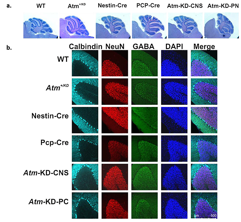
Fig. 5. Normal cerebellar morphology in mice expressing AtmKD in the CNS or in PCs.
Histological analysis of the cerebellum was preformed between ages 1–24 months. Representative images are shown a. Hematoxylin-eosin staining of cerebellar paraffin sections produced from 6-month old mice with different genotypes. b. Immunostaining of different cell populations in cerebellar sections obtained from 6-month old mice. Calbindin D28k (cyan); NeuN (red) highlighting neurons; GABA (green), which marks GABAergic cells; DAPI (blue).

