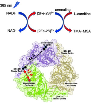
Figure 1.

Schematic of NADH photoactivation coupled with EPR used to monitor the electron transfer from NADH to the Rieske‐type, [2Fe‐2S]2+ in AbCntA variants (e.g. AbCntA; PDB ID: 6Y8J) [9b] and subsequent, carnitine catalysis takes place at the monoFe (where EPR‐silent, ferrous center is converted into high‐spin, ferric species; S=5/2) when the photo‐reduced, AbCntA‐WT sample was annealed at higher temperatures (more details; see Figure 4).
