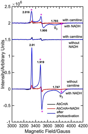
Figure 2.

cw EPR spectra of the as‐isolated “AbCntA‐WT” in the presence and absence of carnitine/NADH following photoexcitation of NADH using blue‐light, 365 nm; (bottom) AbCntA‐WT+NADH in the absence of carnitine; (middle) AbCntA‐WT+carnitine in the absence of NADH; (top) AbCntA‐WT+NADH in the presence of carnitine. The double header arrowed, dotted magenta line indicates that the g 3 component of the Rieske‐type, [2Fe‐2S]+1 EPR signal in the AbCntA‐WT has been shifted towards low magnetic field upon substrate, carnitine binding. The magenta asterisks show the significant EPR line broadening observed in the AbCntA‐WT+NADH+carnitine sample. Conditions; as described in the experimental section in the SI.
