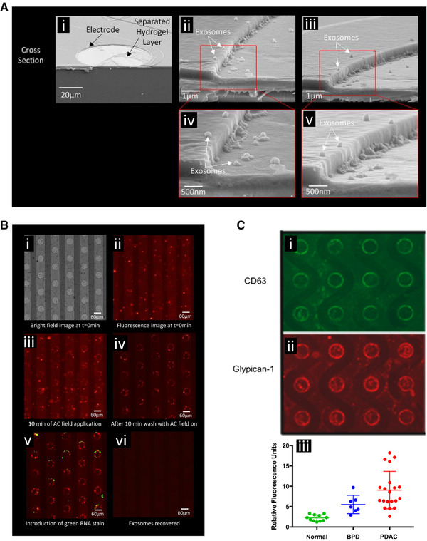
Figure 5.

DEP‐based collection and detection of extracellular vesicles (EVs), including exosomes, and associated cancer biomarkers. (A) Scanning electron microscopy images showing exosome collection around the edges of circular electrodes of the DEP chip where the field strength was the highest. (i) Cell culture derived exosomes were collected from a buffer, the chip freeze dried, and then snapped in half to give cross‐sectional views of the collection. (ii and iii) Further magnification showing exosome collection at the electrode edge. ( iv and v) Further magnification showing individual collected exosomes. Reprinted (adapted) with permission from [52]. Copyright 2017 American Chemical Society. ( B) Collection of fluorescently labeled cell culture derived exosomes spiked into human plasma. (i) Bright field view of the electrode array. (ii) Fluorescent image showing distribution of EVs and exosomes before collection. (iii) Collection of EVs and exosomes around the electrode edges. (iv) The bulk plasma was washed away purifying the collected EVs and exosomes. (v) The collected particles were stained for biomarker content, in this case a green RNA stain was applied. (vi) The DEP field was reversed releasing the collected particles for subsequent off chip analysis. Reprinted with permission from [52]. Copyright 2017 American Chemical Society. (C) Detection of cancer‐associated biomarkers carried by the collected EVs and exosomes. The collected EVs and exosomes stained positively for the (i) CD65 biomarker and (ii) glypican 1 biomarker. (iii) Quantification of glypican 1 levels using DEP collection and immunostaining across different patients showed a significantly higher level of expression in the pancreatic ductal adenocarcinoma (PDAC) patients compared to normal controls. Benign pancreatic disease (BPD) also showed significantly elevated levels. Reprinted with permission from [53]. Copyright 2018 American Chemical Society.
