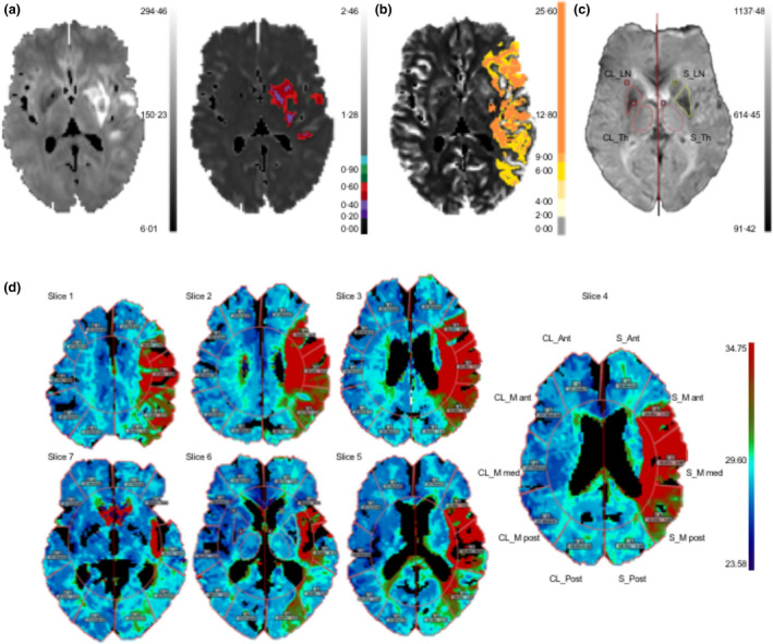
Figure 1.

Workflow for image‐post‐processing with Olea Sphere. (a) Diffusion‐weighted image and automatic segmentation of core volume (right) with apparent diffusion coefficient <600 × 10−6 mm2/s and (b) penumbral volume with a time‐to‐maximum threshold of >6 s. (c) Manual segmentation of the lentiform nucleus (LN) and thalamus (Th) on stroke (S) and contralateral (CL) side. (d) Automatic region of interest segmentation of anterior cerebral artery territory (S_Ant, CL_Ant), middle cerebral artery territory (S_M ant/med/post, CL_M ant/med/post), and posterior cerebral artery territory (S_Post, CL_Post) in seven slices
