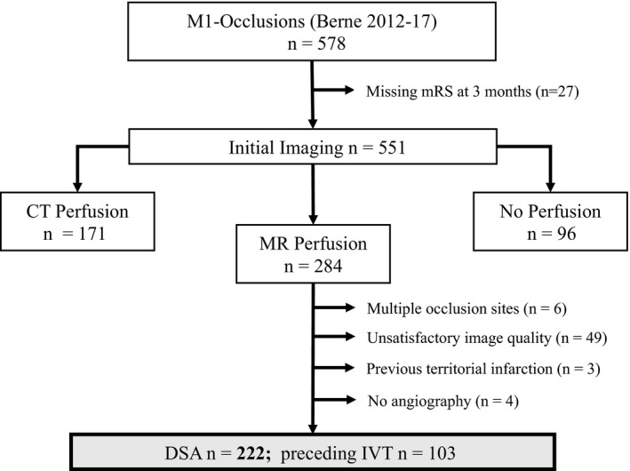
Figure 2.

Flowchart for patient selection. We screened 578 ischemic stroke patients with middle cerebral artery (MCA)‐M1 occlusion and included 222 into the analysis. We excluded 27 patients due to missing modified Rankin Scale (mRS) values at the 3‐month follow‐up time point (n = 551). In this study, we analysed only patients with acute stroke magnetic resonance (MR) imaging including perfusion imaging (n = 284). A total of 62 patients were further excluded either due to insufficient perfusion data quality, for example, because of excessive head motion and other artefacts or missing MR perfusion sequence data (n = 49), additional large vessel occlusion other than extension of the occlusion to the internal carotid artery, or more distal MCA occlusions (n = 6), prior territorial infarction (n = 3), or missing angiography (n = 4). CT, computed tomography; MT, mechanical thrombectomy; DSA, digital subtraction angiography; IVT, intravenous thrombolysis.
