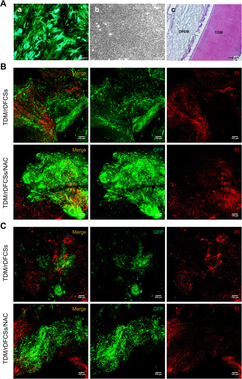
Fig. 4.
Allogeneic transplantation of bioroot composites in a Sprague-Dawley rat bone defect model for 8 weeks. A (a) GFP-labelled rDFCs. A (b) Construction of cell sheets. A (c) Histological section of the TDM scaffold combined with intrinsic fibre three-dimensional dental pulp extracellular matrix, as determined by haematoxylin and eosin (HE) staining. Images of PI staining to detect the survival of grafted cells on B day 1 postimplantation and C day 3 postimplantation. TDM, treated dentin matrix; DPEM, dental pulp extracellular matrix

