
Fig. 3. Reshaping morphogen interpretation with positive or negative feedback.
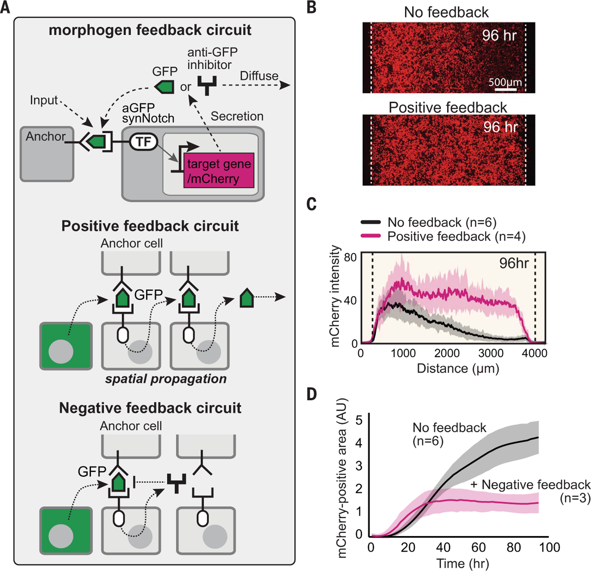
(A) In a positive feedback circuit, GFP morphogen activates receiver cells to induce the secretion of more GFP. In a negative feedback circuit, GFP morphogen induces the expression of antimorphogen inhibitor by receiver cells. TF, transcription factor. (B) Comparison of mCherry output in the body with and without positive feedback at 96 hours (see fig. S7C for time course). The pole has 3 × 10⁴ GFP-secreting cells; the body has a 1.5 × 10⁴ mixture of anchor cells and receiver cells engineered with a positive feedback circuit (50:50 ratio). Images were taken by incucyte system for 4 days (movie S1). (C) Activity gradient profiles at 96 hours, with and without positive feedback. Shaded area shows SD from multiple experiments. (D) The mCherry-positive area (integral of top plots) plotted over time shows that the body with negative feedback reaches steady state faster than it does without feedback. AU, arbitrary units.