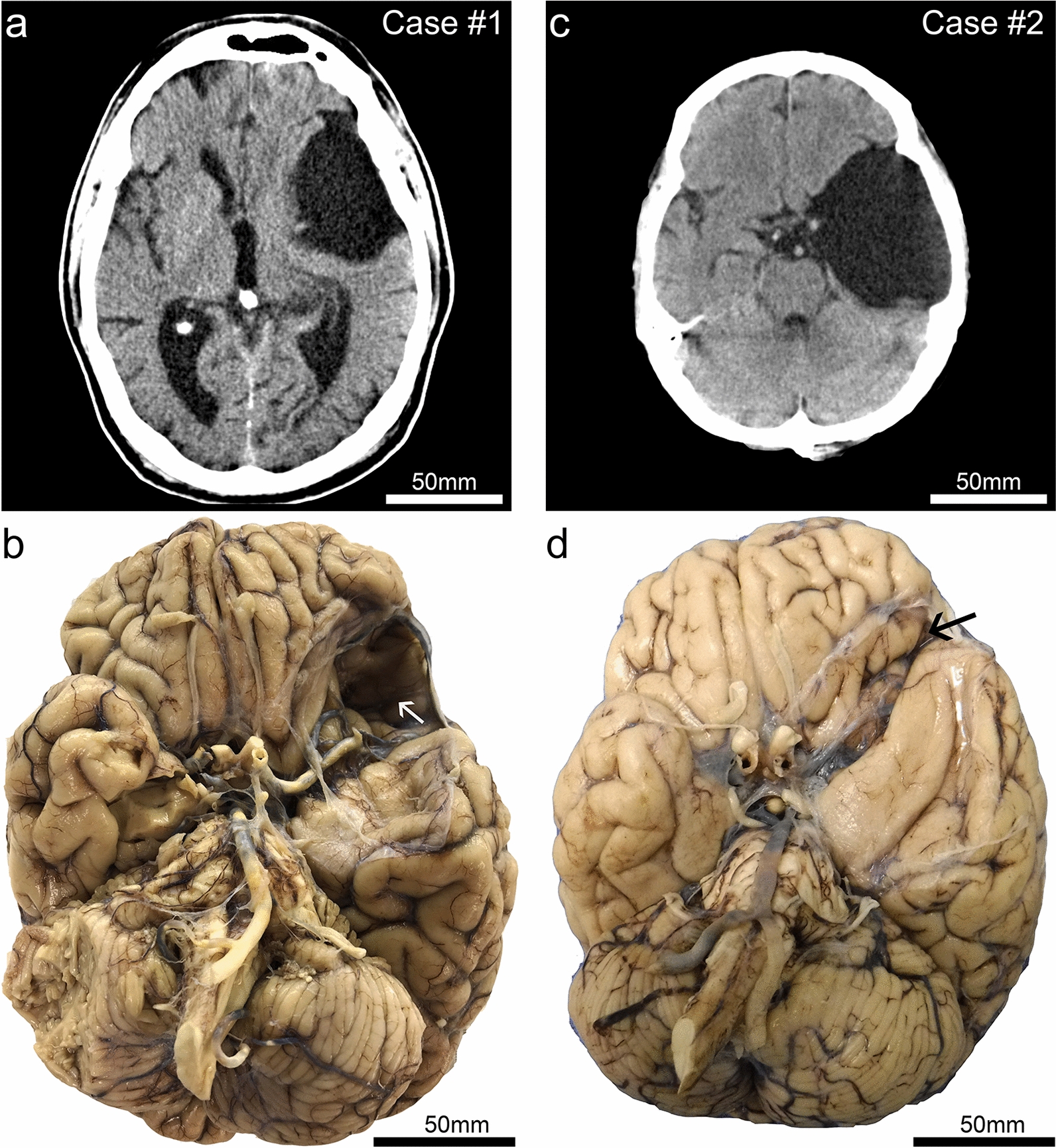
Fig. 1.

Clinical (premortem) computerized tomography (CT) scans, and photographs of brain after autopsy and fixation, in two cases with large arachnoid cysts. Case #1 is a male, 72 years old at death. This CT scan (a) was acquired at age 68. (b) The brain at autopsy following fixation from Case #1 shows the large left-sided arachnoid cyst cavity. Both the anterior temporal lobe and the orbitofrontal frontal lobe are affected by the lesion. Note that on the surface of the frontal cortex within the cyst cavity there is discoloration (white arrow). By contrast, the right side lacks evidence of indentation or contusion in the orbitofrontal cortex. Case #2 is a male, 64 years old at death. The CT scan (c) was acquired at age 56. As seen in the brain at autopsy following fixation (d), the arachnoid cyst is larger in the rostral-caudal axis in Case #2 in comparison to that of Case #1. However, the lesion is shallower—the affected region is smaller in the dorsal–ventral axis, and there was less impact on the orbitofrontal cortex (black arrow) in Case #2 compared to Case #1
