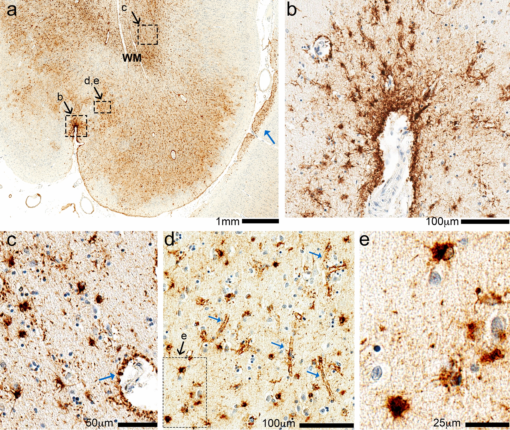
Fig. 3.

Phospho-tau (p-tau) immunohistochemistry in the left fronto-parietal region of Case 1. In the low-magnification photomicrograph (a), the pia mater is near the bottom and white matter (WM) near the top. This is a section of the left fronto-parietal cortex, which is caudal to the region most directly impacted by the arachnoid cyst, and shows widespread p-tau (PHF-1) immunoreactivity in the subpial, gray matter, and white matter regions. In each of these compartments there is prominent staining of p-tau surrounding small blood vessels, and also in cells with morphologic features of astrocytes. Subpial staining is demonstrated in a small sulcus (b) and additional subpial staining is shown at low power in the adjacent gyrus (blue arrow in a). (c) White matter shows p-tau cells with astrocyte morphology, as well as staining around blood vessels resembling astrocyte foot processes (blue arrow). (d) The astrocytic p-tau in gray matter highlight pericapillary staining (blue arrows); the inset shows compact p-tau + cells with astrocyte features (e)
