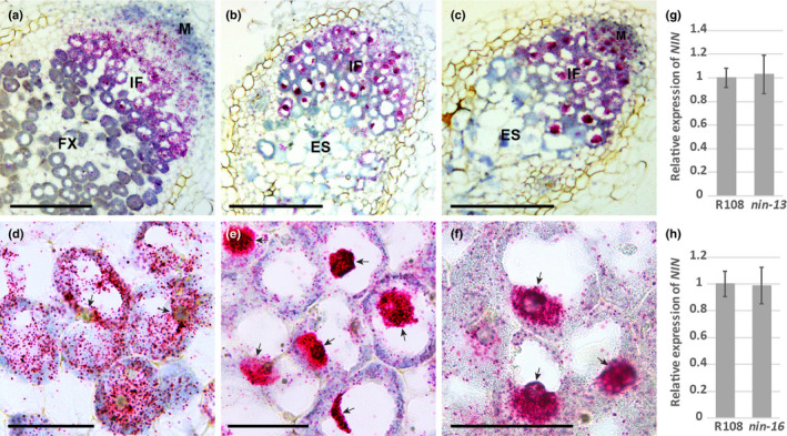
Fig. 5.

NIN mRNA level reduced in the cytoplasm of nodule cells of Medicago nin‐16. RNA in situ localisation of NIN shows that the substantial part of NIN mRNA accumulates in nuclei of nin‐13 (b, e) and nin‐16 nodule cells (c, f), compared with wild‐type (R108) nodule section (a, d). Hybridisation signals are visible as red dots (a–f). Arrows indicate the nuclei (d–f). M, meristem; IF, infection zone; FX, fixation zone; ES, early senescence zone. Quantitative reverse transcription polymerase chain reaction (qRT‐PCR) shows that NIN expression level in wild‐type (R108), nin‐13 and nin‐16 nodules were similar (g, h). Data are means ± SD of three biological replicates. Bars: (a–c) 250 µm; (d–f) 40 µm.
