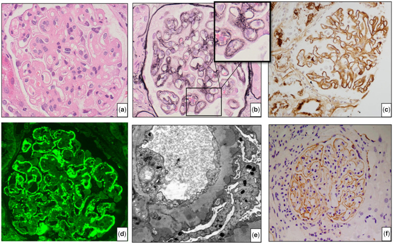
FIGURE 1.
Renal biopsy of Case 15 (MN, HIV and HBV): (A) haematoxylin and eosin staining shows thickened glomerular capillary walls; (B) silver staining demonstrates characteristic spikes and holes along the capillary wall, shown in the inset; (C) IgG staining shows granular staining along the capillary wall; (D) IF for PLA2R shows granular staining along the capillary walls; (E) EM shows typical EDD in the subepithelial area and (F) THSD7A staining of Case 14 (HIN-MN) shows granular staining along the capillary walls.

