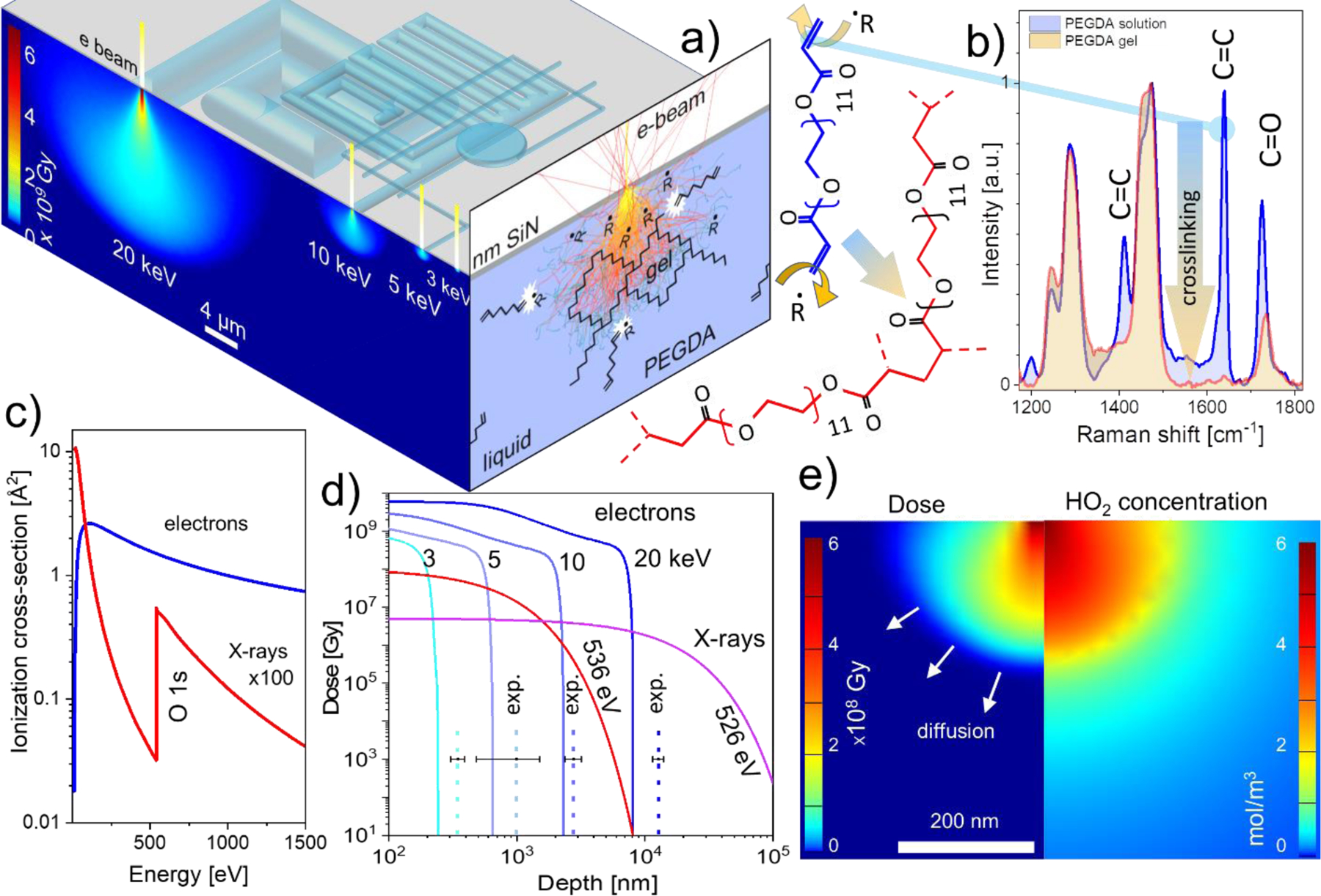
Figure 2.

a) Top facet: the principle of electron beam gelation in liquid polymer solution through 50 nm thick SiN membrane. Lower energy electrons generate smaller excitation volume and therefore finer feature size. Left facet: spatial distribution of the energy deposition by electron beams in water calculated using Monte Carlo simulations. Right facet depicts the concept of direct and indirect (through radiolytic radicals R) PEGDA polymer crosslinking; b) Left: structure of PEGDA 700 (blue) with terminal C=C bonds that are attacked by the beam-generated radicals leading to cross-linking of the PEGDA molecules into polymeric chains (red). Right: vanishing of C=C signatures in PEGDA solution upon irradiation with ca. 106 Gy dose, indicating complete crosslinking of the precursor molecules. c) The comparison of energy dependence of the electron and soft X-ray ionization cross sections of liquid water; d) MC simulated absorbed energy doses as a function of depth for electrons (100 pA current, 1 ms dwell time) and for X-rays (2·109 photons/s and 10 ms dwell time). Dashed vertical lines depict experimentally observed hydrated feature sizes created using the same exposure parameters. Error bars represent standard deviations from three separate experiments. e) Calculated energy deposited distribution for 3 keV 200 pA electron beam (left panel) and the corresponding distribution of hydroperoxyl radical concentration (right panel)
