
Figure 6.
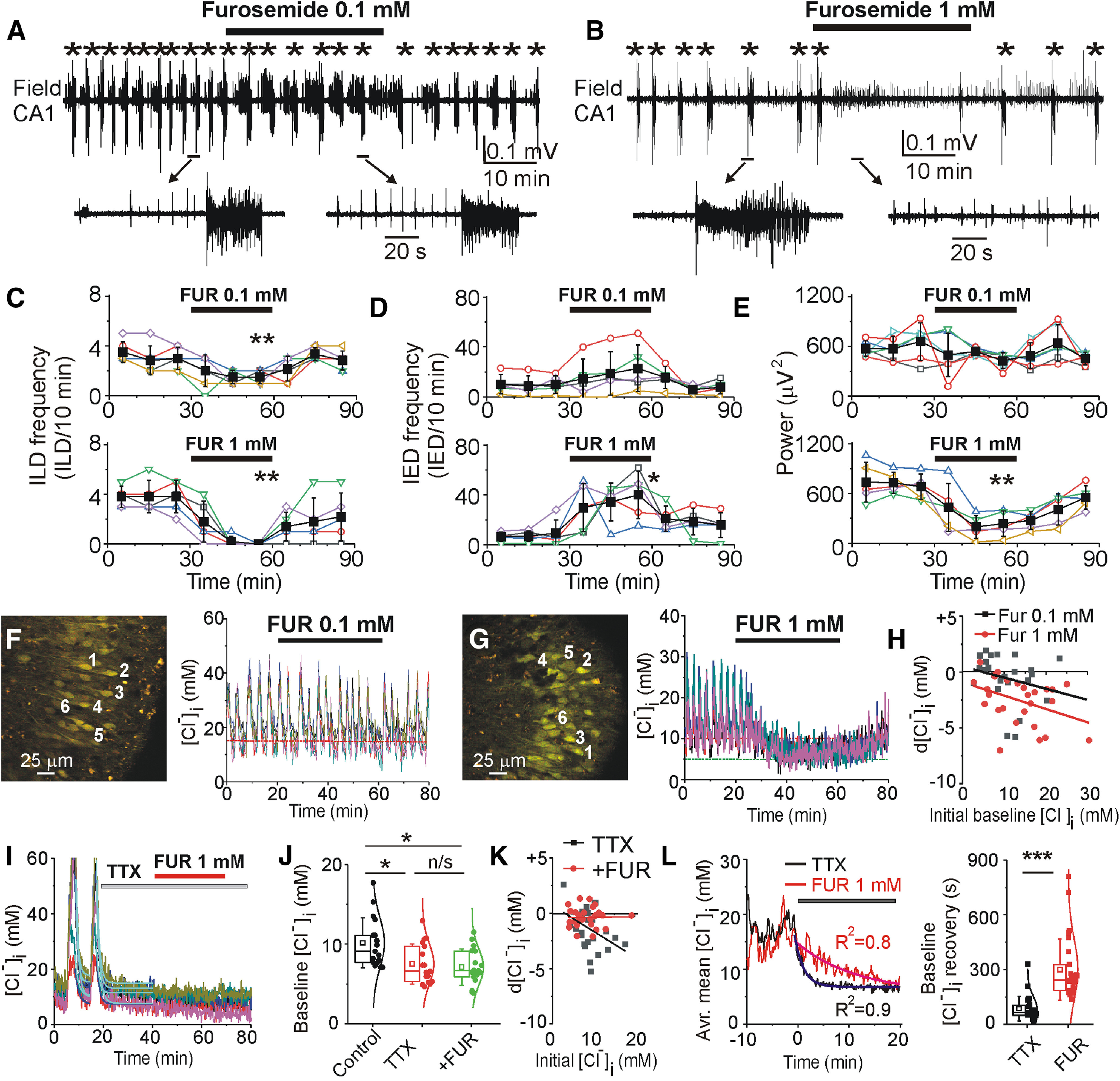
Effects of NKCC1/KCC2 blocker furosemide on recurrent epileptiform discharges and [Cl–]i. A, B, Extracellular field potential recording in the CA1 pyramidal cell layer in the organotypic hippocampal slice at DIV22 before (control), during and after application of furosemide (0.1 and 1 mm for 30 min). C–E, Summary plots of the frequency of ILDs, IEDs, and power of corresponding activity in 10-min windows in individual slice cultures (open symbols). Filled symbols indicate mean ± SD. Furosemide (0.1 mm) reduced the frequency of ILDs and increased the frequency of IEDs. Furosemide (1 mm) abolished recurrent ILDs, increased the frequency of IEDs, and significantly decreased the power of electrical activity (*corresponds to p < 0.05; **p < 0.01, paired sample t test). F–H, Effects of furosemide (0.1 and 1 mm) on [Cl–]i. Furosemide (1 mm) abolished recurrent ILDs and corresponding [Cl–]i transients, and progressively reduced the baseline [Cl–]i. H, Baseline [Cl–]i changes in the presence of 0.1 mm furosemide and 1 mm furosemide as a function of an initial baseline [Cl–]i. I–K, Effects of TTX (1 μm) and furosemide (1 mm) in the presence of TTX on [Cl–]i. TTX rapidly abolished chloride transients, reduced the median baseline [Cl–]i (*p < 0.05 compared with control, Friedman RM ANOVA on ranks, Tukey’s test) and prevented effects of furosemide on [Cl–]i (p > 0.05 compared with TTX). L, Averaged mean [Cl–]i changes before and after applications of TTX (black) or furosemide (red). Data were fitted with exponential fit to calculate decay time of [Cl–]i extrusion during suppression of ILDs. Box (left) + data (right) plots correspond to median (25−75%) [Cl–]i in paired cells (filled circles) and their distribution curves; open squares and whisker range indicate mean ± SD. Furosemide significantly delayed chloride extrusion during suppression of ILDs (***p < 0.001; Mann–Whitney rank-sum test).