Figure 3.
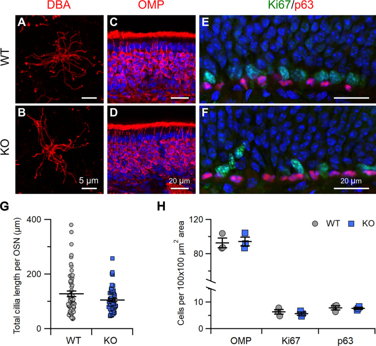
Stoml3 KO mice have grossly normal olfactory epithelium. A, B, Enface view of a whole-mount preparation of olfactory epithelium with OSN cilia labeled by biotinylated DBA detected by streptavidin-Alexa Fluor 594. C–F, Confocal micrographs of coronal sections of the olfactory epithelium from WT and KO mice immunostained for OMP (C, D) or Ki67 and p63 (E, F). Nuclei were stained with DAPI (blue). G, Quantification of the total cilia length per OSN from WT and KO mice (OSNs: n = 61 from 4 WT mice, n = 57 from 4 KO mice). H, Quantification of OMP-immunopositive, Ki67-immunopositive, and p63-immunopositive cells in olfactory epithelium from WT and KO mice.

