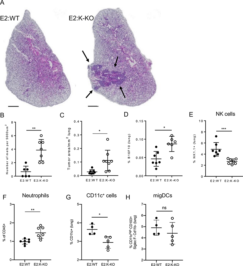
Figure 5. Metastatic cancer is better controlled when nonclassical monocytes are able to patrol.
A) Hematoxylin and eosin (H&E) stained lungs were prepared from E2:WT and E2:K-KO mice i.v. injected with 5×10⁵ B16F10 cells 2 weeks before harvesting. Images represent one biological triplicate. Scale bar 1000 μM. Error bars = 95 % CI. B) The area of total tumor mass over total lung area for each H&E slide analyzed. C) Number of B16F10 metastases quantified per section of H&E slide analyzed. Each point is an individual section from a total of 3 biological replicates. D) E2:WT and E2:K-KO BMT mice were sacrificed 16 hours after B16F10-RFP injection and the frequency of B16F10-RFP cells out of all lung cells was measured between E2:WT and E2:K-KO BMT mice. n = 6–7 mice per group. E) frequency of NK1.1+ cells in lungs of E2:WT compared to E2:K-KO mice. n = 6–7 mice per group. F) frequency of Ly6G+ neutrophils in the vasculature of lungs of E2:WT compared to E2:K-KO mice. n = 6–7 mice per group. G) Frequency of CD11c+ cells in lungs of E2:WT compared to E2:K-KO mice. n = 6–7 mice per group. H) Frequency of migratory dendritic cells (DCs) in lungs of E2:WT versus E2:K-KO mice. Kolmogorov-Smirnov nonparametric test for single comparisons. ns = not significant, *p<0.05, **p<0.01, ***p<0.001.

