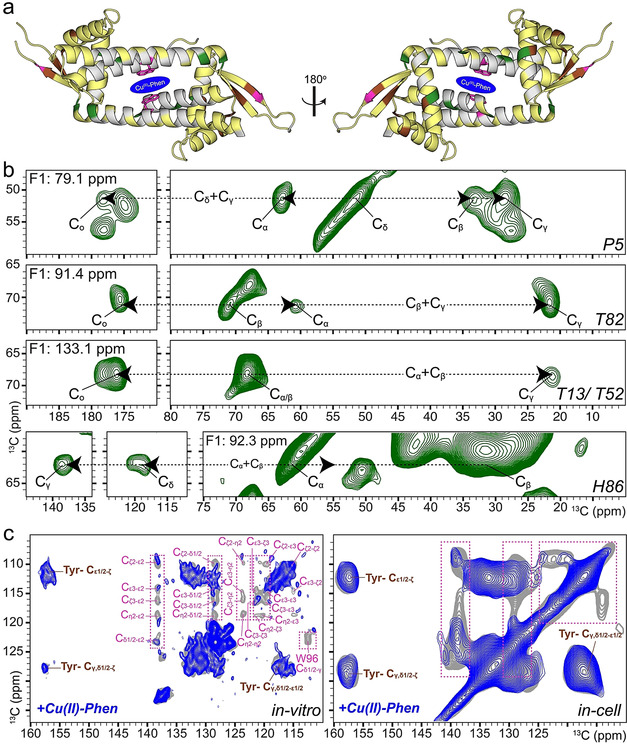
Figure 3.

In vitro and in‐cell NMR studies of the LmrR_A92E/CuII‐Phen artificial metalloenzyme. a) Summary of NMR analysis: Residues used for both solution‐ and solid‐state NMR analysis are plotted on the crystal structure of LmrR (PDB ID: 3F8F) in yellow, green, pink and brown. Green residues were identified in the 3D 13C (DQ‐SQ‐SQ) DNP‐ssNMR spectrum (b). Tryptophan (pink) and tyrosine (brown) residues are used for analysis in (c), in which the selective reduction of NMR signal intensities for Trp aromatic signals confirms proper in‐cell assembly of the ArM. In‐cell experiments were performed in E. coli Lemo21 (DE3).
