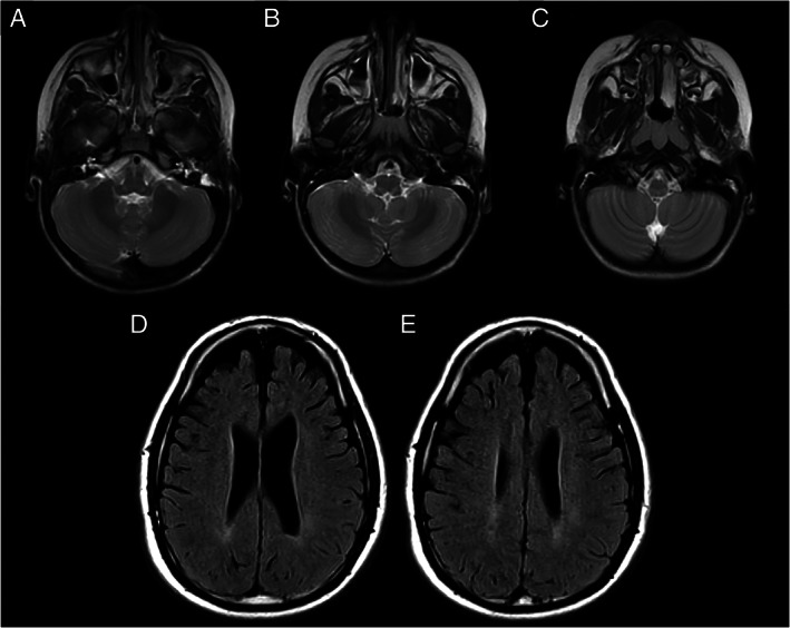
FIGURE 2.

Neuroimaging. (A–C) Brain magnetic resonance imaging (MRI; T2‐axial view) in Patient C‐II‐1 (de novo p.Gly130Arg variant), showing abnormal, focal concentric, symmetric hyperintense signal abnormalities in the posterior brain stem at the bulbomedullary junction. (D, E) Fluid attenuated inversion recovery (FLAIR) MRI images (axial view) in Patient E‐II‐3 (p.Asn32Thr variant), showing bilateral frontal–parietal atrophy and mild hyperintense signal in the posterior periventricular white matter.
