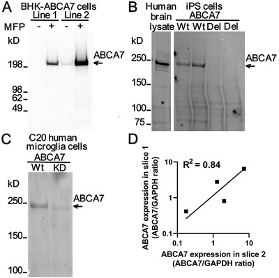
FIGURE 5.

Specificity and repeatability of ABCA7 level measurement using the E11ABCA7 monoclonal antibody. A, Detection of human ABCA7 in two BHK cell lines that express it weakly (line 1) and strongly (line 2) in an MFP‐inducible manner. In line 1, the antibody did not recognize any protein in cells that were not treated with MFP and detected a protein migrating as a single band > 200 kDa in size in cells that were treated with the inducer. In line 2 with strong expression of ABCA7, the antibody detected leaky expression of the protein in uninduced and very strong expression of the protein in MFP‐induced cells. B, Detection of ABCA7 in human brain tissue and human iPS cell lysates. iPS cells either expressed ABCA7 (wild‐type [Wt]) or were deleted in ABCA7 (del). C, Expression of ABCA7 in Wt or ABCA7 knock‐down (KD) human microglial C20 cells as revealed by the ABCA7 antibody. D, Correlation of the ABCA7 levelbetween two independently obtained and processed slices of the hippocampus from four individuals
