Fig 4.

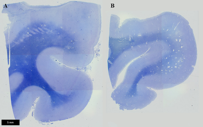
Perivascular dilatation in the cerebrum. In the temporal lobe, enlarged perivascular spaces are observed on sections, stained Klüver–Barrera, from a DM1 patient (B) but not a control individual (A).

Perivascular dilatation in the cerebrum. In the temporal lobe, enlarged perivascular spaces are observed on sections, stained Klüver–Barrera, from a DM1 patient (B) but not a control individual (A).