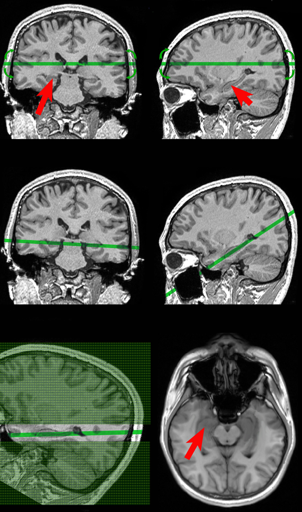
Figure 1.

Selection of oblique hippocampal/temporal cortex plane at the MRI console.
Top: A trained operator with experience in neuroanatomy is presented with coronal & sagittal views of 3D MPRAGE volume. Red arrows indicate the right and the left hippocampus. The operator manipulates the position and the orientation of oblique acquisition plane while monitoring the 3D anatomical alignment (green line and green arrows).
Middle: The ASL exam begins after selecting the optimal plane through both the left and the right hippocampus.
Bottom left: the green area represents tagging, and the difference between slice non-selective (shaded in green) and slice selective slab (not shaded). Note that slice selective slab is 3.5 times thicker than imaged slice.
Bottom right: The resulting balanced steady-state free precession image used to measure CBF. Red arrows indicate the hippocampus.
