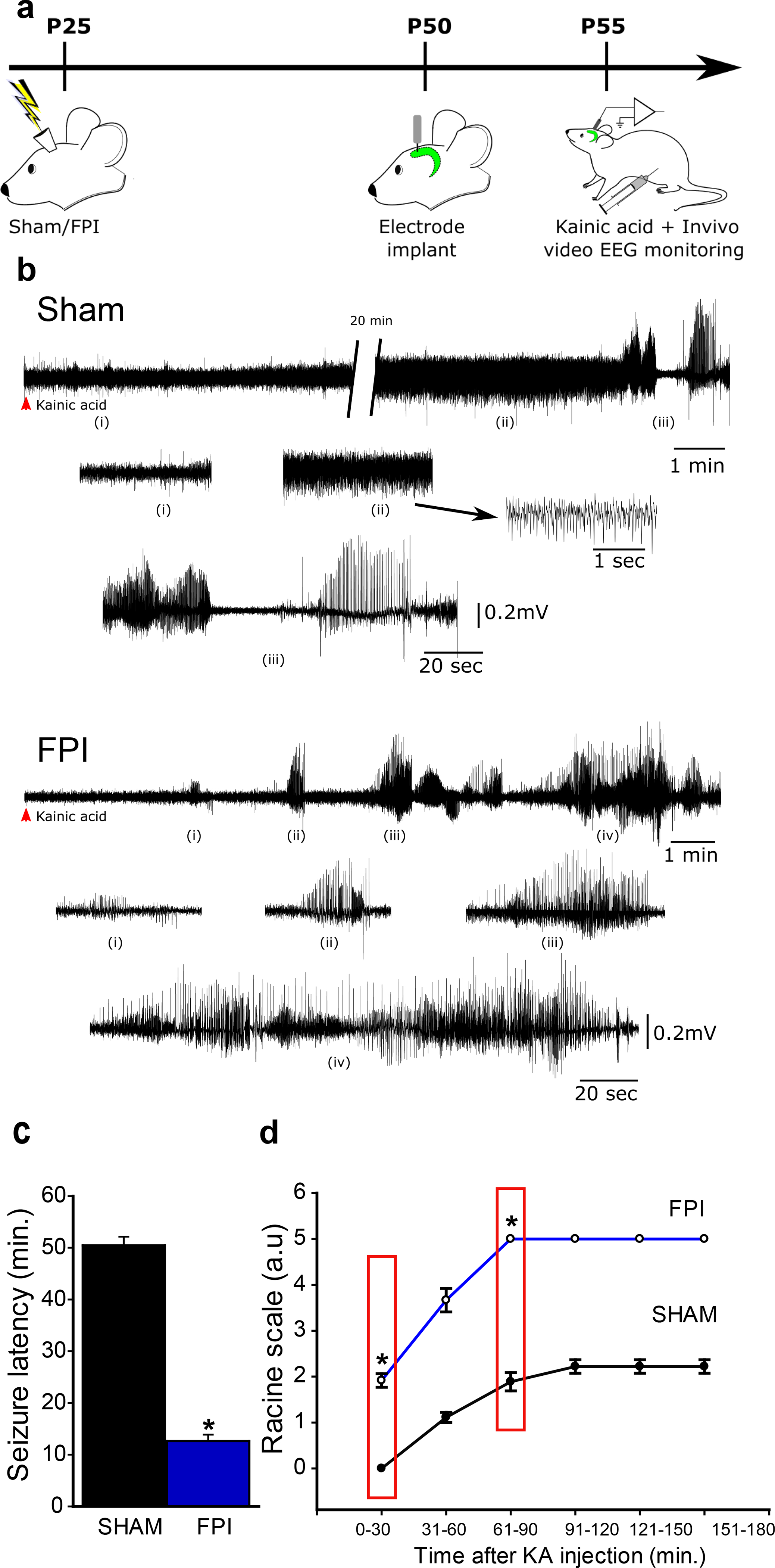
Figure 3. Enhanced susceptibility to chemically evoked seizures after brain injury.

a. Schematic of timeline for in vivo injury followed by electrode implantation and low-does Kainic Acid challenge b. Sample hippocampal depth electrode recordings show evolution of electrographic activity following kainic acid injection in rats one month after sham (above) and FPI. Note that FPI animals develop electrographic seizures early which quickly develops into convulsive seizures (FPI, i-iv). c. Summary of latency to kainic acid induced seizures in rats 1-month after sham or brain injury. d. Summary of progression of seizure severity by Racine’s scale over time in sham (n=9) and FPI (n=12) rats. Boxed areas indicate time points at which statistical comparisons are reported. * indicates p<0.05 by Student’s t-test.
