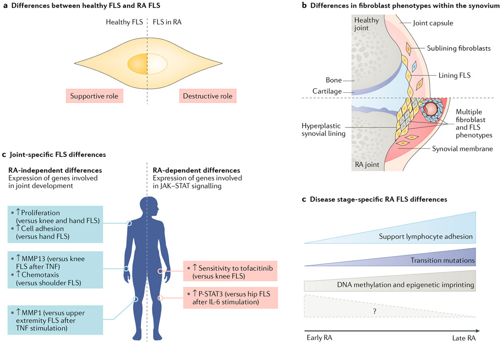
Fig. 3 ∣. Diversity of FLS in RA.
a ∣ Fibroblast-like synoviocytes (FLS) in healthy joints and FLS in joints affected by rheumatoid arthritis (RA) are very different. For example, healthy FLS mainly have a supportive role in the synovium and support structures. RA FLS, on the other hand, have an aggressive, imprinted phenotype and have a destructive role. b ∣ Fibroblasts, including FLS, within the same joint have a variety of phenotypes, especially in RA. These phenotypes might represent true subsets and/or these phenotypes might reflect the local environment in which they reside. c ∣ FLS show spatial heterogeneity with biological differences between various joints. These differences can include RA-independent differences in the expression of genes involved in cell differentiation such as HOX and WNT genes (blue boxes; examples from Frank-Bertoncelj et al.162) or RA-dependent differences in the expression of genes, including genes involved in cytokine signalling through Janus kinase (JAK)–signal transducer and activator of transcription (STAT) (red boxes; examples from Hammaker et al.164). Arrows indicate changes in levels of transcripts or functions for the indicated joint compared with other joints. d ∣ The function of RA FLS varies depending on the stage of disease. FLS in the late stage of RA promote lymphocyte adhesion to endothelial cells and can have genetic alterations. Differential methylation of DNA in RA FLS occurs early in disease but evolves as the disease progresses. Additional studies on FLS in very early RA are needed to dissect some initial pathogenic pathways (shown by the question mark); future omics analyses will hopefully shed light on these processes.

