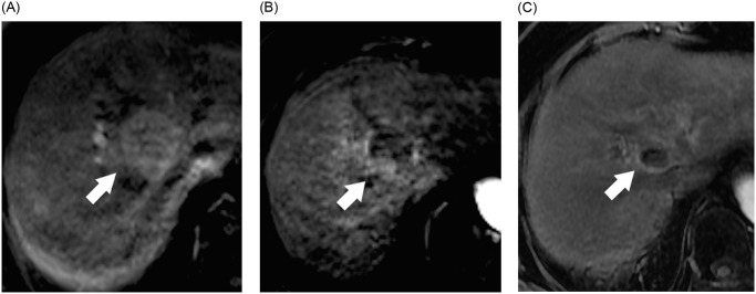
Fig 1. Early tumor regression (ETR) of the hepatocellular carcinoma (HCC) after proton beam therapy (PBT).
74-year-old woman with cirrhosis from chronic hepatitis C virus infection and HCC. MR image obtained before (A), 1 month (B) and 5 months after (C) PBT are shown. (A) Axial T1-weighted image during arterial phase obtained before PBT shows a 3.7 cm HCC in segment 8 (arrow). (B) Axial T1-weighted image during the arterial phase obtained 1 month after PBT shows ETR of the tumor with greater than 50% decrease of diameter of viable tumor (arrow) compared with pretreatment image. (C) Axial T1-weighted image during the arterial phase obtained 5 months after PBT shows lack of enhancement in lesion (arrow); this finding is compatible with complete response.

