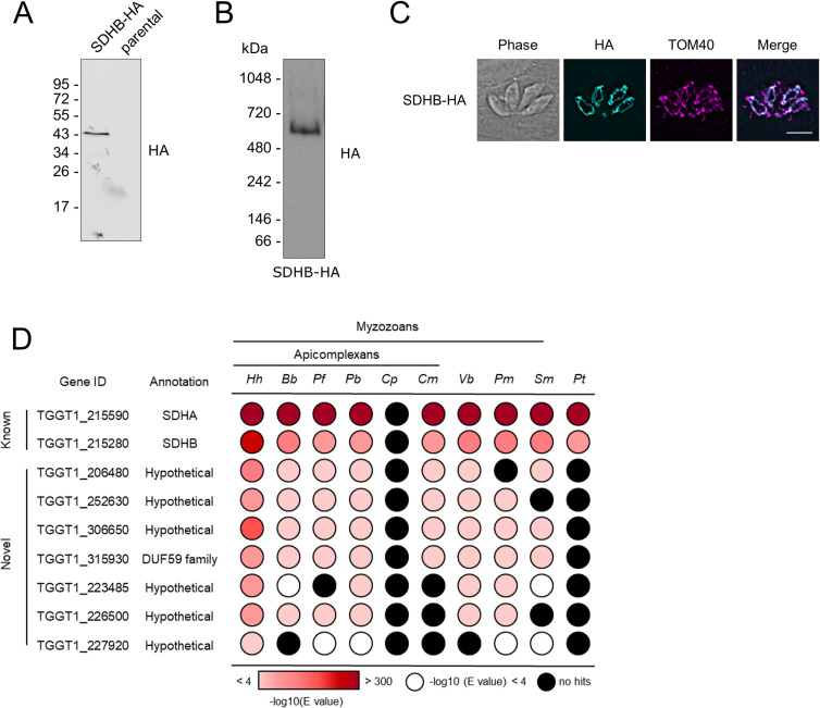
Fig 3. Analysis of putative Toxoplasma complex II subunits.
(A) Immunoblot analysis of complex II subunit SDHB endogenously tagged with an HA epitope. Proteins from total lysate were separated by SDS-PAGE and detected using anti-HA antibodies. Parasites from the parental line (TATIΔku80) were analysed as negative control. (B) Total lysate from SDHB-HA separated by BN-PAGE and immunolabelled with anti-HA antibodies. (C) Immunofluorescence assay with parasites expressing the endogenously HA-tagged SDHB (cyan), showing co-localisation with the mitochondrial marker protein TOM40 [48] (magenta), along with merge and phase. Scale bar is 5 μm. (D) Table showing the previously predicted (known) and complexome identified putative novel (novel) complex II subunits and their homology distribution across key groups. Homology searches were performed using the HMMER tool [57]. Coloured circles refer to the e-value from the HMMER search: white indicates a hit with an e-value above 0.0001, black indicates no hits, and red indicates hits with an e-value below 0.0001, as indicated in the coloured scale. Full data are given in S7 Table. Hh: Hammondia hammondi; Bb: Babesia bovis; Pf: Plasmodium falciparum; Pb: Plasmodium berghei; Cp: Cryptosporidium parvun; Cyryptosporidium muris; Vb: Vitrella brassicaformis; Pm: Perkinsus marinus; Sm: Symbiodinium microadriaticum; Pt: Paramecium tetraurelia.

