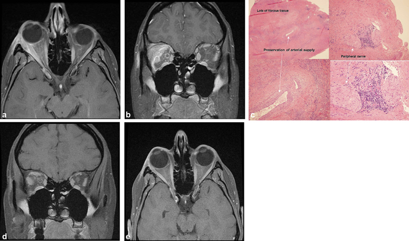
Fig. 1.

( A, B ) Axial and coronal magnetic resonance imaging (MRI), T1 with contrast: the right lacrimal gland and lateral rectus are enlarged and enhance as do the lateral/superior periorbita and optic nerve sheath; stranding of the adjacent orbital fat. ( B ) Coronal MRI, T1 with contrast: the right lacrimal gland and lateral rectus are enlarged and enhance as does the lateral/superior periorbita and the optic nerve sheath. There is also stranding of the adjacent orbital fat. ( C ) Right lacrimal gland. Hematoxylin and eosin (H&E) × 20 upper left, × 100 lower left and upper right, ×200 lower right: note the abundance of fibrous tissue and loss of lacrimal gland architecture with preservation of arterial supply. There is a peripheral nerve with adjacent inflammation which may account for some of the discomfort associated with this condition. ( D, E ) Axial and coronal MRI, T1 with contrast: postoperative scan at 6 months shows mild residual enlargement of right superior and lateral rectus muscles and resection of lacrimal gland mass.
