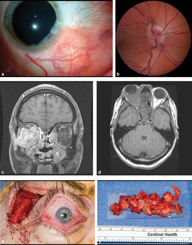
Fig. 2.

( A ) Slit lamp photograph of right cornea showing a pseudopterygium characteristic of healed marginal keratitis from 3:00 through 6:00. ( B ) Fundus photograph of right eye demonstrates swollen optic disk. This resolved following debulking surgery. ( C ) Axial magnetic resonance imaging (MRI) T1 without contrast shows that the right orbit is virtually filled with fibrous tissue. ( D ) Coronal MRI, T1 with contrast: the right medial, inferior and lateral rectus muscles are mildly enlarged and a contrast enhancing infiltrate surrounds these muscles and the optic nerve. There is a small infiltrate in the medial portion of the left orbit. ( E ) Intraoperative photography of a no-bone flap lateral orbitotomy with sutures around each rectus muscle insertion. Gentle traction on suture is transmitted to corresponding muscle belly allowing identification of each encased muscle. All of the palpable retrobulbar tissue was removed through this incision. ( F ) Gross pathology of debulked orbital fibrous tissue.
