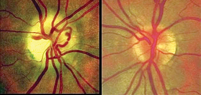
Fig. 7.

Fundus photos demonstrate normal appearance of the left optic nerve head, but right optic atrophy and a prominent cilioretinal shunt vessel in this patient with a history of progressive loss of vision of the right eye found to be secondary to optic nerve sheath meningioma (This image is provided courtesy of Steven A. Newman, MD).
