
Fig. 16.
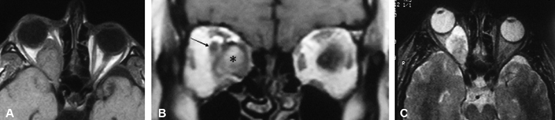
( A ) T1-weighted axial MRI without contrast reveals a large right intraconal mass largely replacing the orbital fat, with posterior extension initially believed to involve the optic canal. ( B ) T1-weighted coronal MRI with contrast without fat suppression delineates the tumor (asterisk) as being distinct from the optic nerve (arrow), which is displaced superolaterally. ( C ) Axial T2-weighted MRI images show a mixed tumor with hyper intense and hypointense signal with sparing at level of the right optic canal. MRI, magnetic resonance imaging.