
Fig. 18.
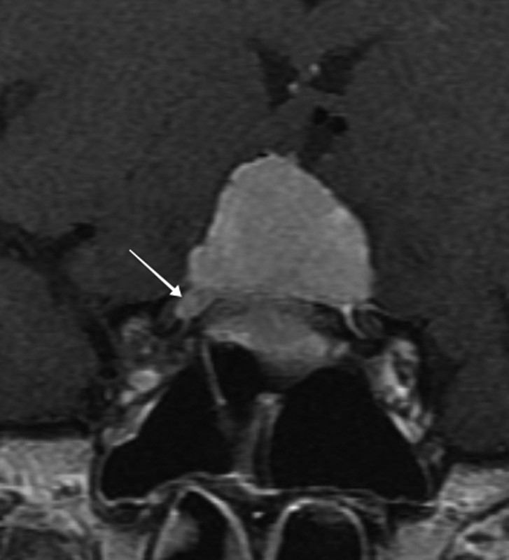
Coronal T1-weighted contrast-enhanced MRI image shows a large dural based planum sphenoidale lesion consistent with a meningioma extending into the right optic canal (arrow). In the setting of visual loss from bilateral optic nerve compression, the patient would benefit from surgical debulking, potentially with a right optic canal decompression and likely subsequent radiotherapy. MRI, magnetic resonance imaging.