
Fig. 5.
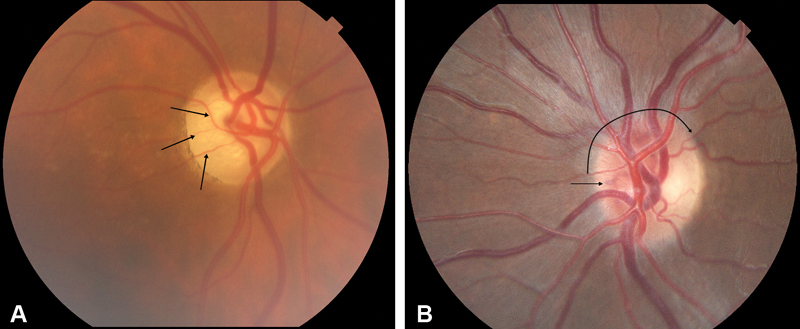
( A ) Right eye fundus photograph from patient with right-sided ONSM, demonstrating moderate temporal optic atrophy with narrowing of segmental components of retinal arteries on the disc (arrows), but no disc swelling nor optociliary shunt vessels seen in the classic triad. This optic atrophy is indistinguishable in this case from optic atrophy caused by other etiology. ( B ) Fundus photograph of left optic disc of a patient with pONSM, mild disc edema (curved arrow) and subtle nasal deep optociliary shunt vessel (arrow). ONSM, optic nerve sheath meningioma; pONSM, pONSM, primary ONSM.