
Fig. 7.
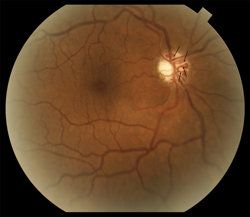
A fundus photograph of the right optic nerve in a patient with pONSM shows prominent optociliary shunt vessels (arrows) and optic atrophy without the presence of known optic disc edema. It is possible disc edema may have regressed prior to observation. pONSM, primary optic nerve sheath meningioma.