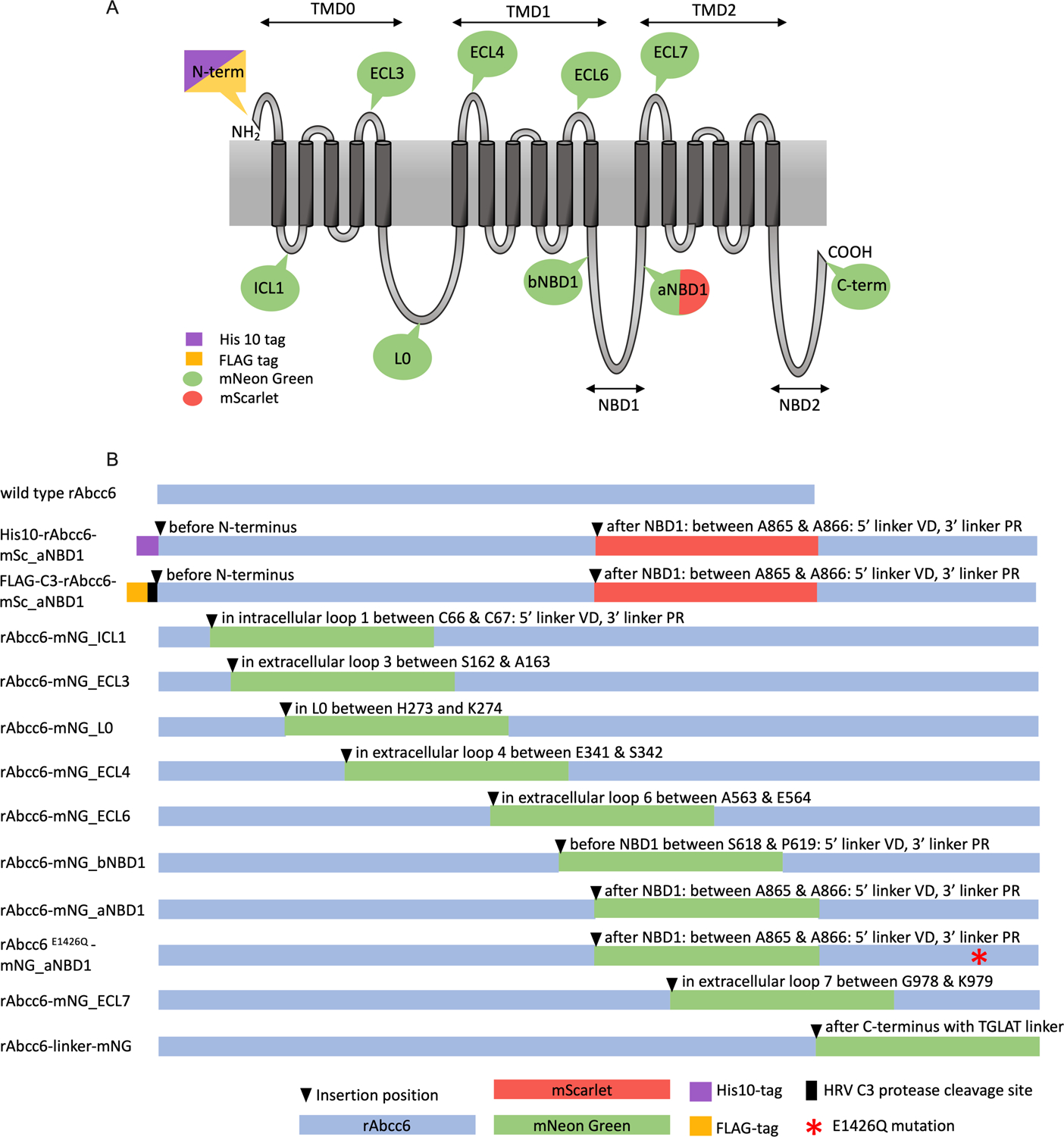
Figure 1. Overview of the rAbcc6 fusion protein constructs.

A: Schematic representation of the membrane topology of rAbcc6 with the locations of the fluorophores and tags. B: Schematic overview of the generated fusion proteins indicating the positions where the mNeonGreen or mScarlet fluorophores were introduced. His10, FLAG and HRV C3 protease cleavage sites are also depicted as is the mutation of the Walker B catalytic glutamate residue (E1426Q).
