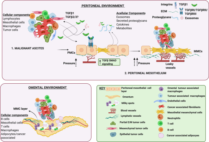
Fig. 3.
Ovarian cancer metastatic environment. The peritoneal and ascites environment are tightly linked to each other as leakage through the peritoneal mesothelium drives malignant ascites accumulation. Malignant ascites is composed tumor cells (in E, M or partial E/M states), either alone or in aggregates composed additionally of immune cells (macrophages, T cells, B cells and neutrophils), fibroblasts, and endothelial cells. Additional non cellular components include multiple cytokines such as TGFβ, exosomes that carry TGFβ, its receptors and also noncoding RNAs, metabolites and proteoglycans that are secreted primarily by the peritoneal mesothelial cells. In the peritoneal mesothelium, TGFβ1 released from tumor cells and CAFs can stimulate TGFβ/SMAD signaling in PMCs driving MMT, that can potentiate vascular changes leading to leakage and altered angiogenesis. Cell aggregates via integrins adhere to MMCs promoting metastasis by ECM degradation and vascular changes. The omental environment supports cell aggregate attachment to the omental MMCs, and growth preferentially near “milky” spots composed of lymphocytes, macrophages, and adipocytes. ECM - Extra cellular matrix, MMT - Mesothelial mesenchymal transition, PMCs - Peritoneal mesothelial cells, MMCs - Mesothelial mesenchymal cells, CAF - cancer associated fibroblasts

