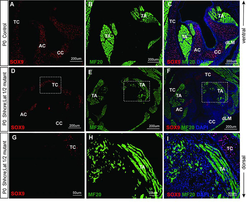
Figure 11: Nuclear YAP in epithelial cells causes mesenchymal defects as noted by distorted laryngeal cartilages and muscle development.
(A-I) Immunofluorescence detection of SOX9 (red), MF-20 (green) and DAPI (blue) in transverse sections of control (A-C) and Shhcre;Lats mutant VF (D-I) at P0. SOX9 (red) staining show SOX9 positive cells in developed laryngeal cartilages and MF-20 (green) staining shows completely developed laryngeal muscles in control VF (A-C). In the mutant larynx, VF show reduced SOX9 staining and defects in development of laryngeal cartilages (D-I) affecting the attachment of the laryngeal muscles. MF-20 staining shows altered structure and bulk of muscles fibers resulting in deformities in thyroarytenoid muscle and the dorsal laryngeal muscles. Primary antibodies are color-coded according to their secondary antibodies, and nuclei are counterstained with DAPI (blue). IF images were taken at 20X, and 40X magnification. Scale bars of 200um and 50um are used in the figure panels. Abbreviations: AC, arytenoid cartilage; CC, cricoid cartilage; TC, thyroid cartilage; EP, epithelium; TA, thyroarytenoid muscle; dLM, dorsal laryngeal muscles.

