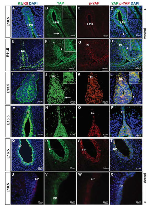
Figure 2: YAP expression in embryonic VF epithelial development.
(A-D) Transverse sections of mouse primitive laryngopharynx at E10.5. IF analysis of K8 (green) (A), total YAP (green) (B), p-YAP (red) (C) and double staining of YAP and p-YAP (yellow) (D). Prominent nuclear YAP in seen in K8 positive progenitors. White solid arrows in the panels of B, D denote nuclear YAP positive cells. These regions are magnified in the same panels in the upper right corners. (E-H) Transverse sections of mouse laryngeal cavity at E11.5. IF analysis of K8 (green) (E), total YAP (green) (F), p-YAP (red) (G) and double staining of YAP and p-YAP (yellow) (H). White solid arrows in the panel of F, H denote nuclear YAP positive cells. These regions are magnified in the same panels in the upper right corners. (I-L) Transverse sections of mouse laryngeal cavity and VF at E13.5. IF analysis of K8 (green) (I), total YAP (green) (J), p-YAP (red) (K) and double staining of YAP and p-YAP (yellow) (L). YAP continuous to be nuclear at this stage and p-YAP is cytoplasmic. White solid arrows in the panels of J, K indicate nuclear YAP positive cells. These regions are magnified in the same panels in the upper right corners. (M-P) Transverse sections of mouse VF at E15.5. IF analysis of K8 (green) (M), total YAP (green) (N), p-YAP (red) (O) and double staining of YAP and p-YAP (yellow) (P). YAP is no longer nuclear in VF epithelial progenitors at this stage. Nucleocytoplasmic shift in YAP localization occurs at E15.5 resulting in total YAP (green) and p-YAP (red) co-localizing together in the cytoplasm. (Q-T) Transverse sections of mouse VF at E16.5. IF analysis of K8 (green) epithelial marker (Q), total YAP (green) (R), p-YAP (red) (S) and double staining of YAP and p-YAP (yellow) (T). (U-X) Transverse sections of mouse VF at E18.5. K5 (red) (U), total YAP (green) (V), p-YAP (red) (W) and double staining of YAP and p-YAP (yellow) (X). YAP is detected only in the cytoplasm where it co-localizes with the p-YAP. Primary antibodies are color-coded according to their secondary antibodies, and nuclei are counterstained with DAPI. IF images were taken at 40X and 60X magnification. Scale bar of 50um and 20um are denoted in figure panels. Abbreviations: LPH, laryngopharynx; EL, epithelial lamina; EP, epithelium.

