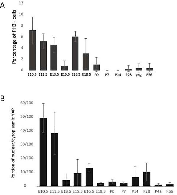
Figure 5: Quantitative assessment of cell proliferation and the portion of nuclear/cytoplasmic YAP positive VF epithelial cells throughout developmental stages.
(A) Quantitative assessment of PH3 positive cells in the VF epithelium throughout the VF developmental stages. (B) The portion of nuclear/cytoplasmic YAP positive cells in the VF epithelium throughout the developmental stages. For each developmental time-point (12 developmental stages in total), three larynges from three different individuals were dissected and analyzed (n=3; 36 WT larynges in total). A General Linear Model procedure with a one-way analysis of variance (ANOVA) determined p< 0.0001 for (A) PH3 and (B) nuclear/cytoplasmic YAP distribution across time points.

