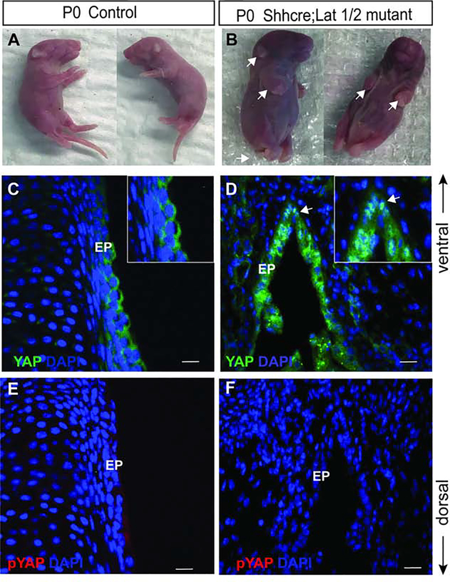
Figure 6: Conditional deletion of Lats1 and Lat2 result in developmental defects in Shhcre;Lats mutants.
(A-B) Whole embryo images of littermate controls at P0 (A) and Shhcre;Lats mutant animals at P0 showed limb, tail and skin defects (white arrows) (B). (C-D) Immunofluorescence detection of YAP (green) and DAPI (blue) in transverse section of VF of control (C) and Shhcre;Lats mutant (D). A white arrow in the panel of D indicates nuclear YAP in VF epithelium. Boxed magnified images in the upper right corners in the panel of C, D show YAP expression in the VF epithelium. (E, F) Immunofluorescence detection of p-YAP (red) in control (E) and Shhcre;Lats mutant (F) showing no p-YAP signal in mutant VF epithelium. Primary antibodies are color-coded according to their secondary antibodies, and nuclei are counterstained with DAPI (blue). IF images were taken at 60X magnification, scale is 20μm. Abbreviations: EP, epithelium.

