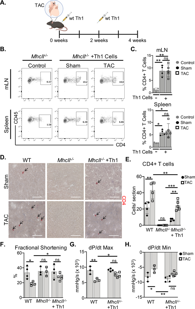
Figure 2. MhcII−/− mice reconstituted with Th1 cells do not develop cardiac dysfunction in response to TAC.
(A) Wt Th1 cells were differentiated in vitro and transferred to MhcII−/− mice via intraperitoneal injection, 2 days and 2 weeks after Sham and TAC. surgery. (B-C) The reconstitution with CD4+ T cells was evaluated in the mediastinal lymph nodes (mLNs) and spleen by FACS, and compared to control MhcII−/− mice that did not receive Th1 cells. (D-E) Frozen LV tissue sections isolated from wt, MhcII−/−, and MhcII−/− mice reconstituted with Th1 cells, 4 weeks after Sham or TAC surgery were used to determine CD4+ T cell LV infiltration. (F) Transthoracic echocardiography was used to measure LV fractional shortening from wt, MhcII−/−, and MhcII−/− mice reconstituted with Th1 cells. (G-H) LV hemodynamic measurements were acquired to determine dP/dt max and dP/dt min as parameters of cardiac contractility and relaxation respectively in MhcII−/− mice reconstituted with Th1 cells compared to untreated wt mice. Scale bars: 100μm. Error bars represent mean ± SD. (* p<0.05, ** p<0.01, *** p<0.001; one-way ANOVA test, two-way ANOVA with two categorical grouping variables).

