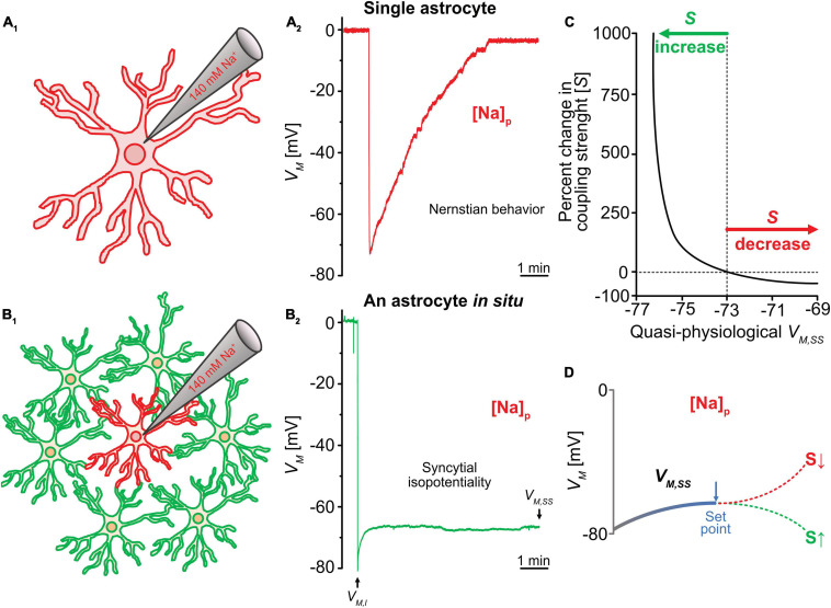
FIGURE 3.
Analysis of syncytial isopotentiality. (A) In a single freshly dissociated astrocyte, the K+ free/Na+ containing electrode solution ([Na+]p) progressively substituted the endogenous K+ content in whole-cell recording, leading to a VM depolarization toward 0 mV as predicated by Nernstian equation. (B) The VM recorded from an astrocyte in situ with the [Na+]p disobeys the Nernstian prediction. Instead, the steady-state VM (VM,SS) maintained at a quasi-physiological level as a result of voltage compensation by the physiological VM of the coupled neighbors. (C) The relationship between VM,SS and coupling strength (S) predicted by computational modeling. (D) According to this modeling shown in (C), the VM,SS can be shifted toward a more hyperpolarizing VM of neighboring astrocytes due to a stronger syncytial coupling (S) or due to a weaker coupling strength (S), the VM,SS toward the GHK predication for the VM of [Na+]p at 0 mV. VM,I stands for initial VM recorded immediately after break-in of cell membrane, reflecting the resting VM of the recorded astrocyte. Figure modified from Kiyoshi et al. (2018).

