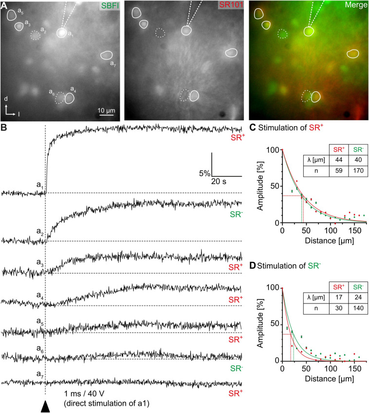
FIGURE 5.
Na+ diffusion in glial networks. (A) Cells in the center of the inferior colliculus can be loaded with the fluorescent Na+ indicator SBFI-AM (left). Astrocytes and bona fide oligodendrocytes can be distinguished by sulforhodamine (SR) 101-labeling. (B) Electrical stimulation results in Na+ load of a single astrocyte (a1). Subsequently, neighboring astrocytes (a3–a5,a7; SR+) and bona fide oligodendrocytes (a2,a6; SR–), show elevated Na+ transients as well. (C,D) Na+ diffusion can be elicited by stimulation of both astrocytes (C) and bona fide oligodendrocytes (D). The amplitude of Na+ transients depends on the distance from the stimulated cell. Figure modified from Wadle et al. (2018).

