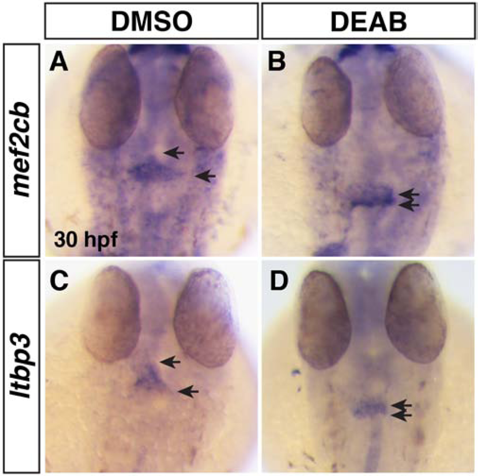
Figure 6. SHF markers of the arterial pole are reduced in RA-deficient embryos.

(A,B) ISH for mef2cb at 30 hpf in DMSO- and DEAB-treated embryos. (C,D) ISH for ltbp3 at 30 hpf in DMSO- and DEAB-treated embryos. Number of embryos examined per condition: mef2cb DMSO-treated n=31; DEAB-treated n=18; for ltbp3 DMSO-treated n=18; DEAB-treated n=16. Views are dorsal with anterior up. Arrows indicate the length of expression at the arterial poles.
