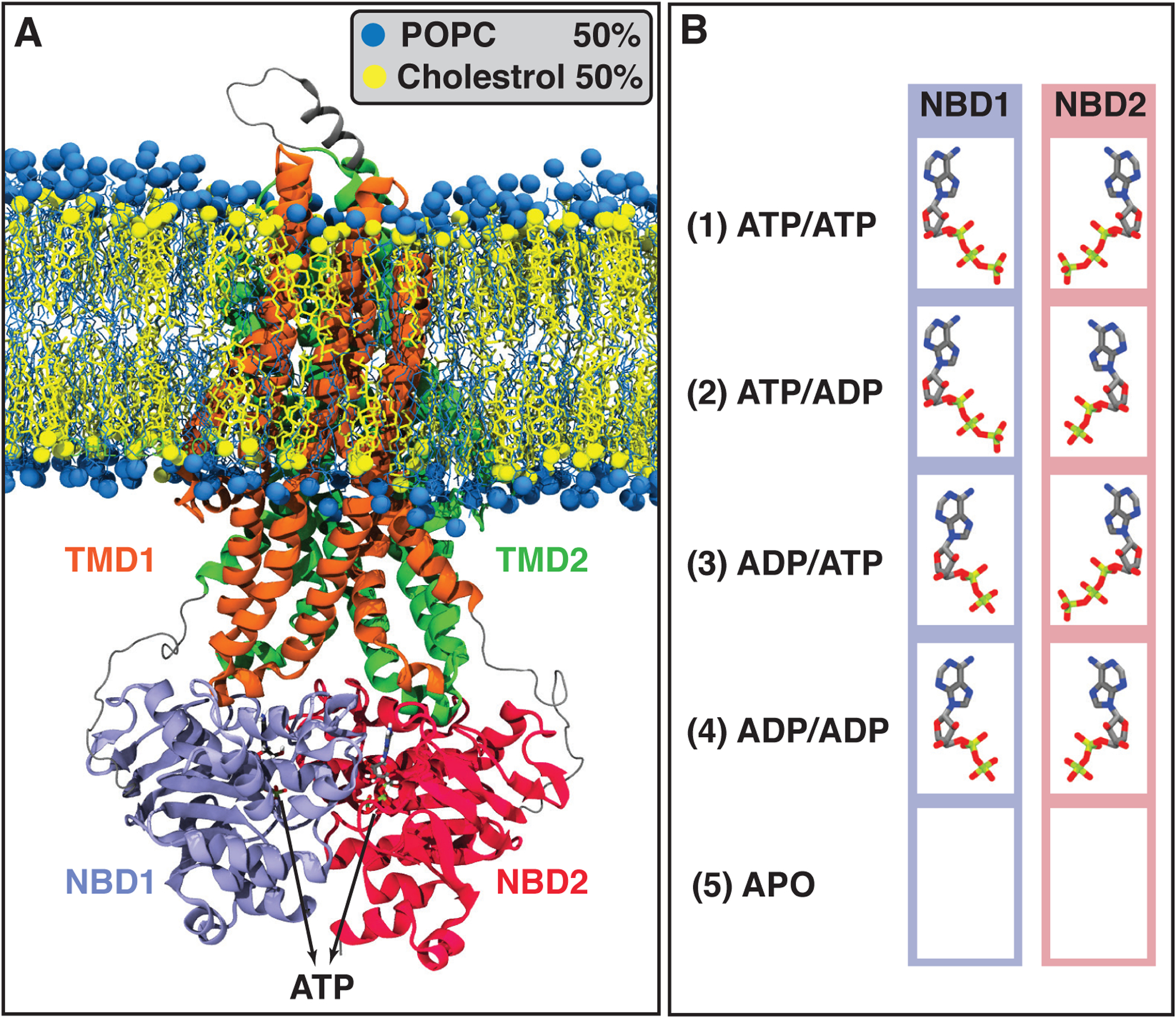
FIGURE 1.

Initial systems configuration including all possible combinations of ATP or ADP bound to the NBDs, as well as the APO (nucleotide-free) system. (A) Pgp ATP-bound structure embedded in a POPC/Cholesterol lipid bilayer (molar ratio 1:1) after 100 ns of MD equilibration. POPC and cholesterol lipids are colored in blue and yellow, respectively. Pgp’s different domains, TMD1, TMD2, NBD1, and NBD2, are colored in orange, green, purple, and red, respectively. (B) The bound states simulated in this study, including all possible combinations of binding nucleotides (ATPs and ADPs), and the APO system which does not include any nucleotide. Each bound state was simulated in three independent simulations.
