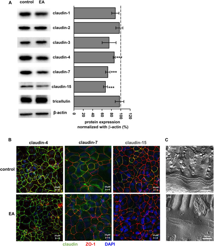
FIGURE 3.
Ellagic acid reduces protein expression of TJ proteins claudin-4, -7 and -15 in Caco-2 monolayers (A) Representative Western blots and densitometry show reduced expression of claudin-4, -7 and -15 in ellagic acid (EA)-challenged monolayers (p***<0.001 vs. control; n = 7–10) (B) Representative micrographs of immunofluorescence staining show claudin-4, claudin-7 and claudin-15 in control and elllagic acid-challenged monolayers (green). ZO-1 served as TJ marker (red), nuclei were DAPI stained (blue) (n = 3 each) (C) Representative freeze-fracture EM micrographs of control and ellagic acid-challenged monolayers show no differences in TJ ultra-structure.

