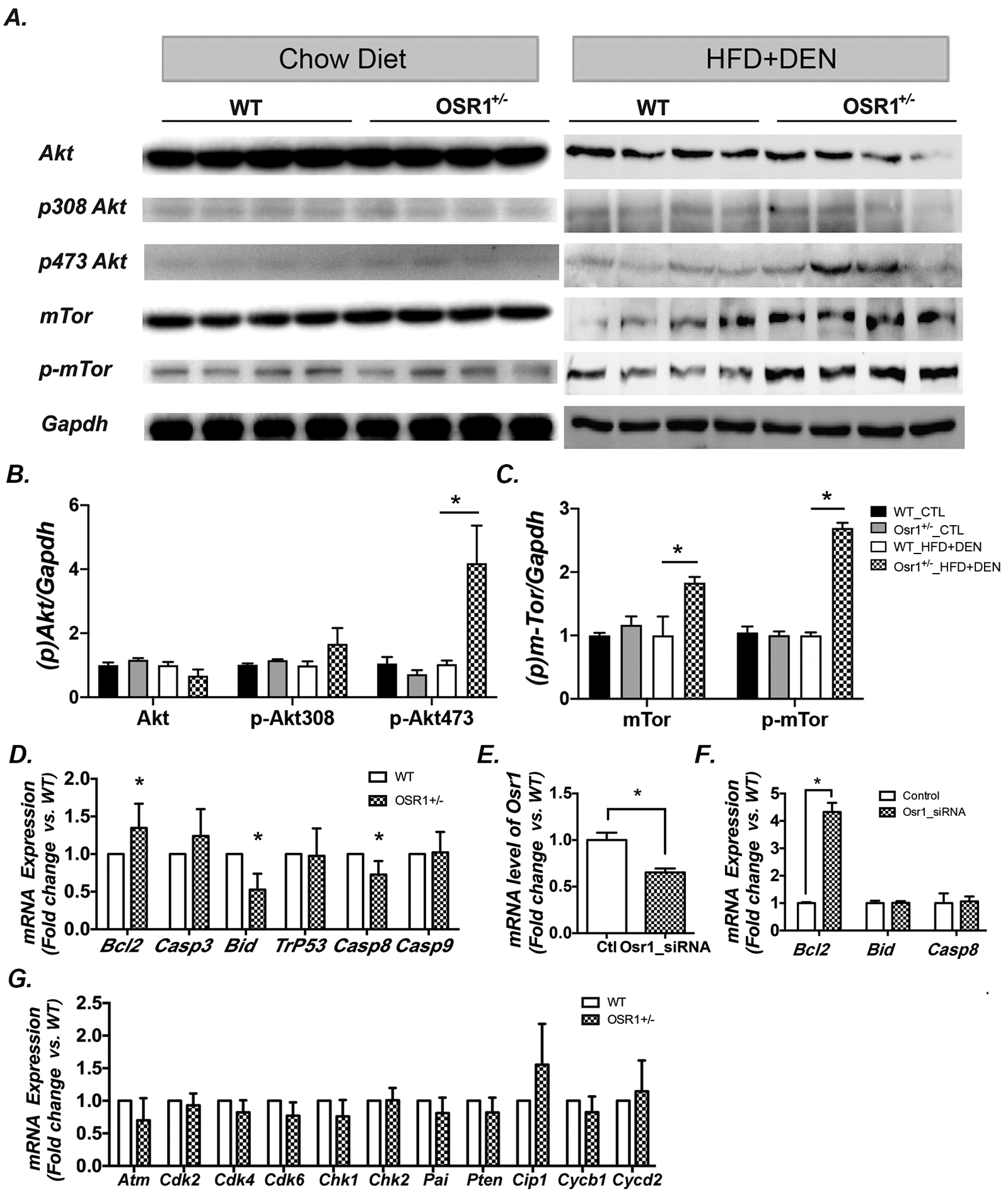
Figure 6.

Osr1+/− mice displayed enhanced hepatic Akt/m-TOR activity and altered expression of apoptotic genes.
(A-C)Western blot of liver tissues showed a higher expression of p-Akt at Ser473 in Osr1+/− mice, but not at Ser308, without effecting basal levels of Akt (6A & B). Higher protein expression of mTOR and p-mTOR in Osr1+/− mice exposed to HFD and DEN were detected by Western blot (6A, C, & D).
(D)Expression of key genes involved in apoptosis was measured by RT-PCR in Osr1+/− and WT livers upon HFD and DEN treatment. Expression of the anti-apoptotic gene, Bcl-2, was up-regulated in Osr1+/− mice, and pro-apoptotic genes, Bid and Casp8, were down-regulated.
(E-F) Knockdown of Osr1 using Osr1-siRNA on HEK293T cells caused an up-regulation of Bcl-2, but not Bid or Casp8.
(G) Expression of key genes involved in cell proliferation was measured by RT-PCR in Osr1+/− and WT liver upon HFD and DEN treatment. None of the cell proliferation related genes were found to have expression alterations between Osr1+/− and WT mice.
Data of figures is presented as Mean ± SE, n=4. * P<0.05 vs. WT group
