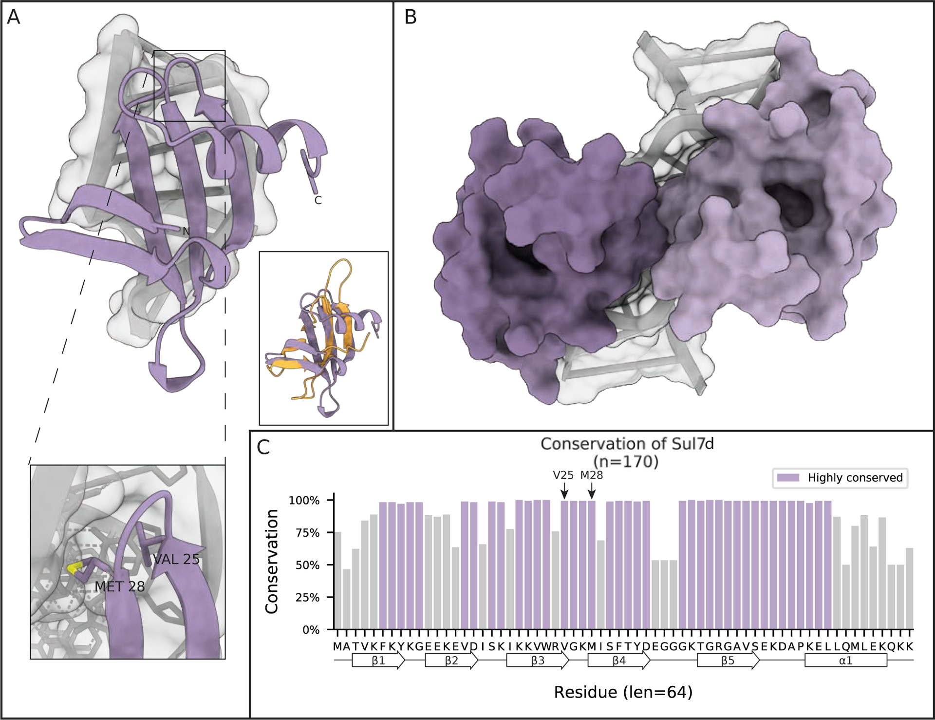
Figure 5: Sul7d kinks DNA.

(A) Crystal structure of the Sul7d-DNA complex showing the kinking of DNA induced by Sul7d from S. acidocaldarius (PDB 1BF4). One inset shows two important residues that intercalate in the DNA double helix in a similar manner as L28 in Cren7 to stabilize the DNA kink. The other inset shows the structure of Cren7 (orange) overlayed onto that of Sul7d (purple). (B) Solution NMR structure of S. solfataricus Sul7d bound to 12 bp of DNA showing the head to head binding mode of Sul7d on DNA. (C) Conservation of Sul7d homologs found in Archaea. Highlighted regions are residues conserved at least one standard deviation more than the mean conservation across the alignment. Secondary structural elements are projected along the residue consensus sequence on the bottom of the plot.
