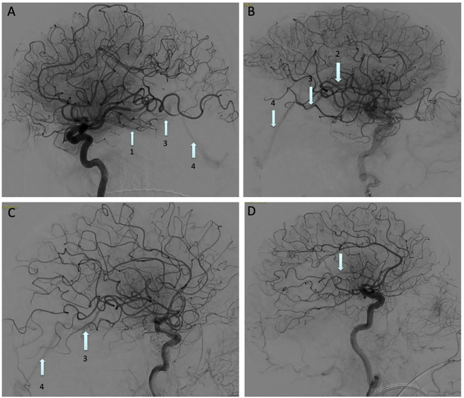
Figure 1.
Digital subtraction angiography images showing an Early Venous Filling (EVF) following recanalization of: (A) M1 occlusion, (B) M2 temporal branch occlusion, (C) M1 occlusion, and (D) Terminal portion of the internal carotid artery occlusion. Basal vein of Rosenthal (arrow 1), internal cerebral vein (arrow 2), great vein of Galen (arrow 3), straight sinus (arrow 4).

